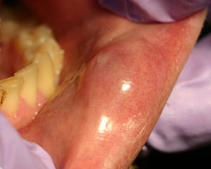
Mucocoele
- A mucococele (also mucocele) is the accumulation of mucus in either the connective tissue of within a salivary duct
- The term encompasses both mucus extravasation phenomenon and mucous retention cyst
Epidemiology[edit | edit source]
Clinical Features[edit | edit source]

- Usually presents as recurrent swelling with intermittent mucous discharge (patients commonly report "drainage and re-filling")
- Commonly there is a history of trauma to the site but this is not always the case
- May develop at any site, but common locations are:[1]
- Lower labial mucosa (81.9%)
- Floor of mouth (5.8%) ← known as a ranula
- Ventral tongue (5.0%)
- Buccal mucosa (4.8%)
- Rare in upper lip
- Clinically appears translucent or bluish in colour (∵ of mucous contents)
- Transillumination is present if the lesion is large enough
- Palpation is usually soft and can feel fluctuant
Differential Diagnosis[edit | edit source]
- Be suspicious of an alternative diagnosis at less common sites (e.g. in upper lip, tumours of minor salivary glands are more common)
Aetiology and Pathogenesis[edit | edit source]

- Not true cysts because there is no epithelial lining (technically, they are polyps)
- Mucous extravasation phenomenon
- Extravasation mucoceles are caused by a leaking of fluid from surrounding tissue ducts or acini
- Usually secondary to trauma causing a rupture of the ducts
- Mucus retention cyst
- Retention mucoceles are formed by dilation of the duct secondary to its obstruction (sialolith or dense mucosa)
Investigations[edit | edit source]
- Diagnosis is made on histology and clinical examination
Histology[edit | edit source]

- Mucin surrounded by granulation tissue
- As inflammation is usually also present neutrophils and foamy histiocytes are also identified
Management[edit | edit source]
- Surgical excision of the cyst and associated minor salivary gland (usually under local anaesthetic)
- Key to avoiding recurrence is to eliminate the adjacent surrounding glandular acini and removing the lesion down to the muscle layer
- Aspiration does not lead to a lasting benefit as the salivary glands quickly refill the mucocoele
- Some reports of using cryotherapy and laser therapy
Prognosis and Complications[edit | edit source]
- Lesions can recur (different rates reported by ~3%)[2]
Follow-up[edit | edit source]
- No routine follow-up needed
- Usually suitable for results to be presented to patient using remote consultation (telephone or results in post)
References[edit | edit source]
- ↑ 1.0 1.1 1.2 Chi AC, Lambert III PR, Richardson MS, Neville BW. Oral mucoceles: a clinicopathologic review of 1,824 cases, including unusual variants. Journal of Oral and Maxillofacial Surgery. 2011 Apr 1;69(4):1086-93.
- ↑ Yamasoba T, Tayama N, Syoji M, Fukuta M. Clinicostatistical study of lower lip mucoceles. Head & neck. 1990 Jul;12(4):316-20.