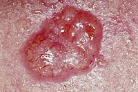
Basal Cell Carcinoma
Commonest form of skin cancer
Slow growing tumour affecting the basal cells of the skin
Can locally invade but rarely metastasises
Can sometimes be referred to by laymen as "rodent ulcer"
Epidemiology
- Accounts for 80% of non-melanoma skin cancers
- 80% of cases occur in the head and neck [1]
- Marked geographical variation (commonest in Europe, Australia and USA)
- Much more common in fair-skinned individuals with a family history
- Up to 30% of caucasians will develop BCCs in their lifetime [1]
- Incidence increases in those >50 years old
- Multiple BCCs in younger patients is suggestive of nevoid basal-cell carcinoma syndrome (Gorlin-Goltz Syndrome)
- ♀ > ♂
Clinical Features
- Most commonly affects sun exposed areas
- Different subtypes exist which tend to occur at different anatomical locations (see below for main types)
- Pigmented subtype also exists (~7% of cases)[2]
Differential Diagnosis
- Squamous Cell Carcinoma
- Malignant melanoma (pigmented basal cell carcinoma)
- Melanocytic naevi (pigmented basal cell carcinoma)
- Bowen's disease (especially superficial basal cell carcinoma)
- Psoriasis (superficial)
- Eczema (superficial)
- Sebaceous hyperplasia
- Molluscum contagiosum
- Appendygeal tumours
Aetiology and Pathogenesis
- Risk factors
- Ultraviolet (UV) light exposure from sun/sun-beds, especially in childhood [3]
- History of previous skin cancers
- Genetic predisposition/association with fair skin type
- Immunosuppression (post-transplant, anti-TNF drugs [infliximab], haematological malignancies)
- Nevoid basal-cell carcinoma syndrome (Gorlin-Goltz Syndrome)
- Xeroderma pigmentosum
- Pathogenesis
Investigations
Diagnosis is usually clinical
In cases where there is uncertainty an incisional or punch biopsy may be performed
Imaging
CT or MRI imaging may be used for extensive lesions to delineate any involvement in adjacent structures
Management
- Management
Prognosis and Complications
- Prognosis and Complications
References
- ↑ 1.0 1.1 Wong CS, Strange RC, Lear JT. Basal cell carcinoma. Bmj. 2003 Oct 2;327(7418):794-8.
- ↑ Maloney ME, Jones DB, Sexton FM. Pigmented basal cell carcinoma: investigation of 70 cases. Journal of the American Academy of Dermatology. 1992 Jul 1;27(1):74-8.
- ↑ Corona R, Dogliotti E, D'Errico M, Sera F, Iavarone I, Baliva G, Chinni LM, Gobello T, Mazzanti C, Puddu P, Pasquini P. Risk factors for basal cell carcinoma in a Mediterranean population: role of recreational sun exposure early in life. Archives of dermatology. 2001 Sep 1;137(9):1162-8.


